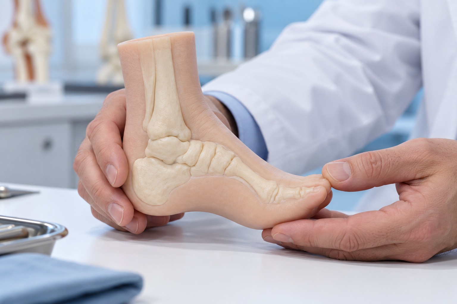
A baby with a convex, rocker-bottom foot may have congenital vertical talus, a structural deformity that differs from clubfoot and flexible flatfoot. By the end, you will know which signs matter, how clinicians confirm the diagnosis, what early casting involves, and why treatment changes with flexibility, age, recurrence, and associated conditions.
Key takeaways
- A persistent rocker-bottom foot needs prompt assessment by a paediatric orthopaedic specialist.
- Examination and weight-bearing or positional X-rays help confirm talonavicular dislocation.
- Early treatment usually uses serial manipulation, casting, and Achilles tendon lengthening.
- Rigid, recurrent, or previously treated deformities may require surgery and longer follow-up.
What Congenital Vertical Talus Looks Like—and How It Differs from Other Foot Deformities
Congenital vertical talus is a rigid deformity in which the talus stays nearly vertical and the navicular is displaced upward and outward from the talar head, creating a persistent talonavicular dislocation. The foot has a convex sole—the characteristic rocker-bottom foot—with the forefoot dorsiflexed and abducted and the hindfoot in equinus and valgus.
The appearance alone cannot confirm the diagnosis. A clinician examines how much the foot can be corrected and uses properly positioned radiographs, including a lateral view and a forced plantar-flexion lateral view.
In true congenital vertical talus, the talonavicular dislocation remains; reduction on the stress view points toward a flexible oblique talus and changes treatment planning.
| Condition | What parents may notice | Key structural difference |
|---|---|---|
| Congenital vertical talus | Rigid rocker-bottom shape; forefoot points upward | Persistent talonavicular dislocation with hindfoot equinus and valgus |
| Clubfoot | Sole turns inward; heel appears small and drawn up | Cavus, forefoot adduction, hindfoot varus, and ankle equinus—not a convex sole |
| Metatarsus adductus | Toes and front half of foot curve inward | Forefoot adduction with a comparatively normal heel and hindfoot |
| Positional deformity | Foot looks turned or bent but improves with gentle movement | Flexible molding deformity without fixed joint dislocation |
| Oblique talus | Flat-looking foot with some rocker shape | Talonavicular alignment improves during forced plantar flexion |
A foot that looks less curved is not necessarily corrected; persistent joint malalignment can remain.
When the Deformity Appears and Why Early Assessment Matters
The deformity is usually visible as a congenital foot deformity at birth, although its practical effect becomes clearer when a child begins to bear weight. The sole has a rocker-bottom contour, and the ankle and midfoot cannot realign normally as the child stands.
A rigid foot can cause difficulty standing and walking because the heel, ankle, and forefoot do not share load in a stable position. As the child grows, pressure along the abnormal plantar surface can produce calluses and shoe fitting problems, pain, poor walking mechanics, and functional limitation.
Early assessment matters even if the infant seems comfortable. Infant joints and soft tissues are more adaptable, while delayed evaluation allows stiffness, pressure-related symptoms, and a harder-to-correct gait pattern to develop. Observation alone is not appropriate for a confirmed rigid deformity.
An apparently isolated foot does not rule out a broader diagnosis. Clinicians commonly perform a neurologic examination and review for:
- Arthrogryposis or cerebral palsy
- Spina bifida
- Chromosomal, connective-tissue, or other syndromic conditions
These neuromuscular and genetic conditions can affect treatment response and increase the risk of residual or recurrent deformity, so follow-up must continue after the initial correction.
How Clinicians Confirm Congenital Vertical Talus
Diagnosis rests on physical examination plus properly positioned radiographs. The examiner looks for a stiff rocker-bottom contour, forefoot dorsiflexion and abduction, hindfoot equinus or valgus, and a forefoot that does not passively realign when gently plantar-flexed. In infants, incomplete bone ossification makes the examination especially important.
| Assessment | Rigid vertical talus | Flexible oblique talus |
|---|---|---|
| Forced plantar-flexion lateral radiograph | The talonavicular dislocation persists while the foot is pushed downward. | The talonavicular joint reduces or moves substantially toward normal alignment. |
| Lateral radiograph | The talus remains nearly vertical, with abnormal talocalcaneal alignment and talo-first-metatarsal alignment. | These relationships improve because the deformity is partially reducible. |
Clinicians obtain weight-bearing foot radiographs when the child can stand. For an infant, they use simulated weight-bearing positioning, including anteroposterior and lateral views. A forced plantar-flexion lateral radiograph is the decisive stress view: persistent dislocation supports congenital vertical talus, while reduction points toward oblique talus and changes treatment planning.
Parents should bring:
- Copies of every prior foot radiograph and the written reports
- Referral letters and records of birth, developmental, neurologic, or genetic evaluations
- A list of diagnoses, medicines, and previous casting or treatment
- Short videos showing standing or walking, if the child is old enough
- Shoes, braces, or casts that clinicians need to examine
What Early Treatment Usually Involves for an Infant
Treatment starts with gentle correction, not forceful straightening. The aim is to bring the talonavicular joint back into alignment and create a plantigrade, flexible foot rather than merely reduce the rocker-bottom appearance.
1. Begin serial manipulation and casting, usually with a reverse-Ponseti technique. The clinician gradually plantar-flexes and adducts the forefoot while supporting the talar head, then applies a long-leg cast to hold each gain. The cast must not be tightened over a swollen or poorly perfused foot.
2. Return for cast changes every one to two weeks. At each visit, the clinician removes the cast, checks skin and circulation, reassesses talonavicular alignment, repeats the manipulation, and applies a new cast. Several rounds may improve flexibility, but a rigid dislocation often does not remain reduced with casting alone.
3. Add a limited procedure when the foot remains rigid or the talonavicular joint will not stay reduced. This commonly combines percutaneous or mini-open Achilles tenotomy and talonavicular reduction, with temporary pin fixation when needed. Achilles tendon lengthening addresses persistent equinus; the reduction restores the relationship between the talus and navicular.
4. Apply another cast after the procedure, then transition to bracing once healing permits. Follow-up checks the joint position, hindfoot alignment, ankle and subtalar movement, and whether the foot remains painless and plantigrade. Open release is reserved for a deformity that cannot be corrected or held through this less-invasive sequence.
How Treatment Changes with Flexibility, Age, Recurrence, and Prior Treatment
Flexibility determines how much correction a child needs. A flexible oblique talus may realign with manipulation and casting, while a rigid congenital vertical talus keeps the talonavicular joint dislocated during forced plantar-flexion.
| Option | What it means | When it applies |
|---|---|---|
| Casting | Serial manipulation and casts hold the foot toward a plantigrade position | Flexible or partially reducible deformity, and as preparation for surgery |
| Limited soft-tissue surgery | Achilles tenotomy, talonavicular reduction, temporary pin fixation, casting, then bracing | Rigid deformity diagnosed in infancy or early childhood when casting alone will not maintain reduction |
| Open release and tendon proceduresで | Wider release of tight joints and tendons to obtain or maintain reduction | Older, severe, untreated, or resistant deformity when minimally invasive treatment fails |
| Osteotomy | Bone is cut and repositioned to correct persistent alignment | Residual or recurrent deformity in an older child after growth, soft-tissue, or joint correction is assessed |
An untreated rigid deformity is rarely managed by observation alone. Older children have stiffer joints and tighter tissues, so forcing correction can damage the skin or create pressure points; surgeons may stage correction rather than perform one extensive operation.
Recurrence is more likely with neuromuscular or syndromic conditions. Follow-up should check talonavicular alignment, foot flexibility, gait, pain, shoe fit, skin pressure, brace use, and whether the child is losing correction.
About The Bone & Joints - Advance Ortho Care can help families compare casting, limited surgery, and osteotomy when examination and radiographs show that the initial correction is not holding.
Related services
Osteotomy and deformity correction Osteotomy and deformity correction Dr. Darshan Shinagne, located on Kolshet Road, Thane, provides advanced osteotomy surgery treatments for patients with joint deformities, arthritis,... View service → |
Paediatric orthopaedics treatment and management Paediatric orthopaedics treatment and management Dr. Darshan Shinagne, based on Kolshet Road, Thane, provides specialized paediatric orthopaedic care for children with bone, joint, and muscle... View service → |
Frequently asked questions
What does congenital vertical talus look like?
It creates a rigid rocker-bottom foot with a convex sole, dorsiflexed and abducted forefoot, and hindfoot equinus and valgus.
When does congenital vertical talus appear, and why does early assessment matter?
It is present at birth. Early assessment identifies the deformity before stiffness, skin pressure, walking problems, and treatment complexity increase.
How do clinicians confirm congenital vertical talus?
Clinicians examine foot flexibility and obtain positional or weight-bearing X-rays to assess talar alignment and persistent talonavicular dislocation.
What early treatment does an infant usually need?
Treatment commonly starts with serial manipulation and casting, followed by procedures such as Achilles tendon lengthening when indicated.
How does treatment change with age, flexibility, recurrence, or prior treatment?
Flexible deformities may respond to less extensive correction, while older, rigid, recurrent, or previously treated feet may need surgery and closer follow-up.
Related service
Foot and Ankle Surgery Dr. Darshan Shinagne, located on Kolshet Road, Thane, offers advanced foot and ankle surgery treatments for various orthopedic conditions and... View service → |