A child with an outward bow in the lower leg, a minor-trauma fracture, or a tibia that will not unite needs more than routine fracture care. By the end, you will understand how congenital pseudarthrosis is identified, how neurofibromatosis type 1 changes evaluation, and how specialists choose between protection, reconstruction, fixation, and long-term follow-up.
Key takeaways
- A dysplastic tibial segment prevents normal bone union after fracture.
- Tibial bowing increases fracture risk and can worsen leg deformity.
- X-rays, comparison views, and MRI reveal the affected bone and soft tissues.
- Reconstruction requires prolonged protection after surgery to maintain union.
What makes congenital pseudarthrosis of the tibia different from an ordinary fracture problem?
Congenital pseudarthrosis of the tibia is failure of the tibial shaft to form a solid union because a dysplastic, biologically inactive segment develops, usually in the distal or distal-middle third.
Its narrowed canal, tapered or sclerotic bone ends, abnormal periosteum, and poor local blood supply make it fundamentally different from a delayed union or nonunion after fracture.
| Condition | What distinguishes it |
|---|---|
| Congenital pseudarthrosis of the tibia | Dysplastic, poorly vascular bone with a persistent gap or fragile union |
| Fibrous dysplasia | An intramedullary bone lesion, not a congenital pseudarthrosis segment |
| Infection | Requires evidence of an infectious process, such as compatible clinical, laboratory, or operative findings |
| Malunited fracture | The bone has united, but in the wrong position |
Neurofibromatosis type 1 (NF1) is the main recognised association: approximately 40–50% of children with congenital pseudarthrosis have NF1, while others are idiopathic or have rarer causes. Crawford classification describes the spectrum from bowing with preserved cortex to visible pseudarthrosis and severe dysplasia.
It helps communicate severity, but classification alone does not predict surgical success; bone quality, age, deformity, fracture status, and fixation strategy also matter.
A narrowed, biologically inactive segment cannot be treated like an ordinary break. The surgeon usually removes abnormal tissue and sclerotic bone until viable bleeding bone appears, then adds bone graft and rigid fixation to address both the biological and mechanical problems.
How does a bowed tibia lead to fracture and progressive leg deformity?
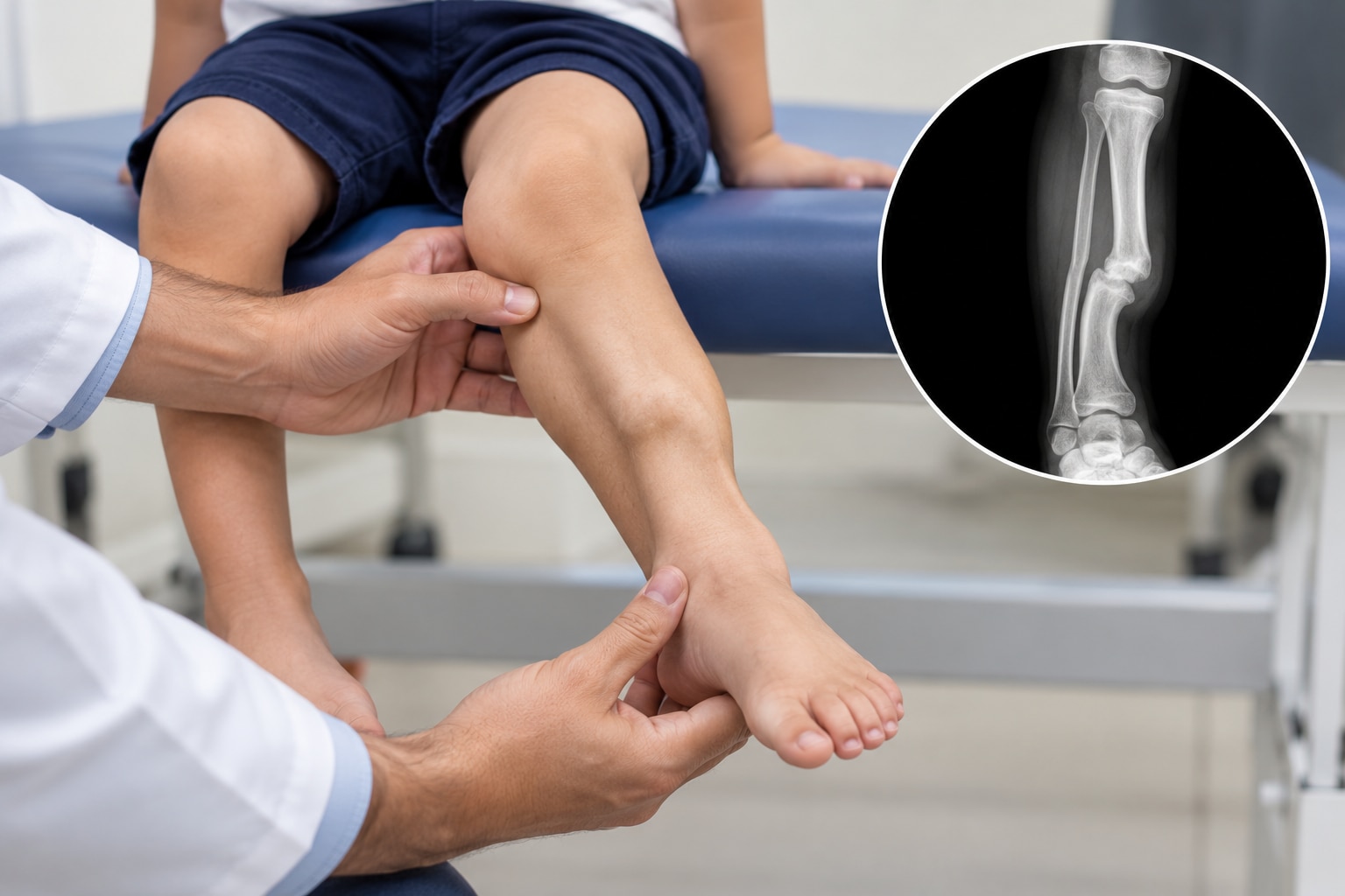
Anterolateral tibial bowing is often visible in infancy, with the lower leg curving forward and outward and a narrowed tibial segment at the apex. This poorly formed area concentrates bending stress, so ordinary walking or minor trauma can exceed the bone’s strength before an obvious break appears.
The problem commonly worsens after walking begins. A tibial fracture may cause pain, but a painless fracture or a progressively unstable leg is the first sign in some children. Recurrent fracture, ankle deformity, and leg-length difference can follow as the tibia bends, breaks, and fails to heal normally.
Associated fibular dysplasia or fibular pseudarthrosis adds a separate source of instability. It can pull the ankle into valgus, produce lateral ankle instability, and accelerate progressive deformity.
Assessment must examine more than the tibial gap:
- Inspect both tibia and fibula for bowing, narrowing, fracture, or pseudarthrosis.
- Check the distal tibial physis, ankle mechanics, alignment, and leg-length difference.
- Record skin condition, pain, instability, and changes after walking begins.
| Structure | What to assess | Why it matters |
|---|---|---|
| Tibia | Bow apex, narrow segment, fracture, alignment | Identifies concentrated stress and failure risk |
| Fibula | Dysplasia or pseudarthrosis | Explains ankle valgus and lateral instability |
| Ankle and physis | Mechanics, growth plate, limb length | Detects deformity that tibial treatment alone can miss |
Which examinations and scans confirm the diagnosis and show its severity?
The diagnosis is confirmed by examination and imaging, not by a single symptom. Examine the skin for café-au-lait macules and other neurofibromatosis type 1 (NF1) features; ask about family history; and inspect the leg and ankle for bowing, swelling, scars, and deformity. Assess pain, skin quality, instability, limb length, and alignment.
NF1 signs can appear over time, so anterolateral bowing or pseudarthrosis warrants paediatric and genetic evaluation when findings or family history support it, even after an incomplete initial examination.
When the child can cooperate, obtain standing long-leg radiographs or a full-length tibia radiograph, with dedicated ankle and knee views.
| Modality | What clinicians assess | When it adds value |
|---|---|---|
| Plain radiographs | Bone-end shape, cortical narrowing, sclerosis, gap size, deformity angle, shortening, fracture status, and whether the fracture is active or healed | First-line diagnosis, severity assessment, alignment, and follow-up |
| MRI | Abnormal bone ends, soft-tissue and marrow changes, and signs suggesting infection | Use when radiographs leave uncertainty or infection is suspected |
| CT | Complex three-dimensional bone anatomy and a potential graft site | Use when plain films cannot define the anatomy adequately |
Radiographs also help describe the Crawford spectrum, from bowing with preserved cortex to visible pseudarthrosis and severe dysplasia. That classification describes severity but does not predict surgical success; bone quality, age, deformity, fracture status, and fixation strategy still matter.
When is observation or bracing reasonable, and when does a child need reconstruction?
Observation is reasonable in a very young child with bowing but no established pseudarthrosis, limited deformity, and stable radiographs. Use serial clinical examinations and imaging; marked bowing, a narrow tibial segment, or radiographic progression increases concern.
A custom brace can protect a vulnerable bowed tibia before fracture in selected children. Bracing does not reliably correct the dysplasia or unite an established pseudarthrosis, so worsening alignment or a fracture changes the decision toward surgical reconstruction.
Once pseudarthrosis is present, surgery must address both biology and mechanics: excise abnormal tissue and sclerotic bone until viable bleeding bone appears, add an autologous bone graft, and provide rigid stabilization. Fixation options have different costs.
| Option | Main advantage | Main limitation |
|---|---|---|
| Telescoping intramedullary rod | Supports the tibia as the child grows and avoids an external frame | Difficult to insert or maintain in a very small, narrow canal |
| Plate | Strong, precise correction and straightforward positioning | Requires a suitable bone segment and can irritate soft tissues |
| Circular external fixation | Powerful compression and correction of complex deformity | Pin-site care, frame management, and joint stiffness |
| Combined construct | Adds stability when one method is insufficient | More extensive surgery and hardware management |
Staged reconstruction may be needed when bone loss, deformity, or soft-tissue problems prevent one definitive procedure. A vascularized fibular graft supplies living bone for extensive loss or repeated failed union, but requires microsurgical expertise and carries donor-site risks.
BMP is generally an off-label, non-routine adjunct supported mainly by small case series. It cannot replace sound resection, grafting, stabilization, and mechanical protection.
What does recovery require after union, and how can families reduce the risk of failure?
An X-ray showing union does not end treatment. Refracture remains a recognised risk before skeletal maturity, after intramedullary rod removal, or while the regenerated tibia is still narrow. Long-term follow-up protects against a healed bone failing during growth.
Repeat radiographs should track more than the union line:
| Follow-up priority | What to check | Why it matters |
|---|---|---|
| Tibial structure | Diameter and new narrowing | A thin tibia remains vulnerable to refracture |
| Alignment and length | Bowing, leg-length difference, and recurrent deformity | Growth can reveal worsening asymmetry |
| Ankle and growth | Ankle mechanics, ankle valgus, and distal tibial growth | Fibular instability can shift ankle loading |
| Implant and fibula | Hardware condition and fibular stability | Loosening or fibular pseudarthrosis can destabilise the leg |
Ask the surgical team:
- How much dysplastic bone will be removed?
- What fixation will fit the child’s canal?
- Which graft source will be used?
- How long will protection continue?
- Which signs indicate infection, hardware failure, recurrent bowing, or refracture?
A paediatric orthopaedic service experienced in limb reconstruction should coordinate care, with genetics or NF1 assessment when indicated.
Families can use About The Bone & Joints - Advance Ortho Care in Thane for an orthopaedic assessment and to discuss referral to a specialised paediatric limb-reconstruction team or a second opinion before major surgery or after failed union.
Related services
Osteotomy and deformity correction Osteotomy and deformity correction Dr. Darshan Shinagne, located on Kolshet Road, Thane, provides advanced osteotomy surgery treatments for patients with joint deformities, arthritis,... View service → |
Fracture Management and Treatment Fracture Management and Treatment Dr. Darshan Shinagne, practicing on Kolshet Road, Thane, provides expert fracture repair treatment for patients with bone injuries caused by... View service → |
Frequently asked questions
What makes congenital pseudarthrosis of the tibia different from an ordinary fracture problem?
The tibia contains a dysplastic, biologically inactive segment that cannot form a solid union normally, so treatment addresses abnormal bone biology as well as the fracture.
How does a bowed tibia lead to fracture and progressive leg deformity?
Bowing concentrates mechanical stress through the weakened distal tibia. A fracture can then shorten, angulate, and worsen the existing deformity as the child grows.
Which examinations and scans confirm congenital pseudarthrosis of the tibia?
Clinical examination assesses bowing, limb length, skin changes, and function. Full-length standing or non-weight-bearing X-rays show alignment and the pseudarthrosis; MRI can assess abnormal tissue and nearby soft structures.
When is observation or bracing reasonable?
Observation or bracing may suit a child with a stable, unfractured tibia and limited deformity. Close follow-up is needed to detect fracture, increasing bowing, or functional problems.
When does a child need reconstruction, and what does recovery require?
Reconstruction is considered for fracture, progressive deformity, shortening, pain, or failure of conservative care. Recovery involves prolonged immobilisation or protected weight-bearing, repeated imaging, and follow-up after union to reduce refracture risk.
Related service
External fixator application External fixator application is a specialized orthopedic procedure used to stabilize complex fractures, severe injuries, and certain bone... View service → |