An elbow that angles outward when straight may reflect normal variation, a congenital difference, or a problem that developed after an elbow fracture. By the end, you will know which symptoms require prompt review, how clinicians confirm the cause, when observation is reasonable, and what corrective treatment involves.
Key takeaways
- Previous elbow fractures are a common cause of cubitus valgus in children.
- Pain, weakness, numbness, or a visible worsening angle warrants an examination.
- Exercises cannot straighten a fixed bony carrying-angle deformity.
- Surgery is considered for substantial deformity, nerve symptoms, or functional problems.
What Is Cubitus Valgus, and What Causes It in Children?
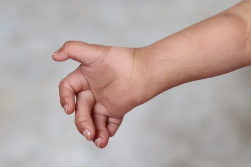
The cubitus valgus definition is an excessive outward forearm angle when the elbow is fully straight. The forearm points farther away from the body than expected relative to the upper arm. The normal carrying angle in children varies with age, sex, side, and measurement method, but is often roughly 5–15 degrees.
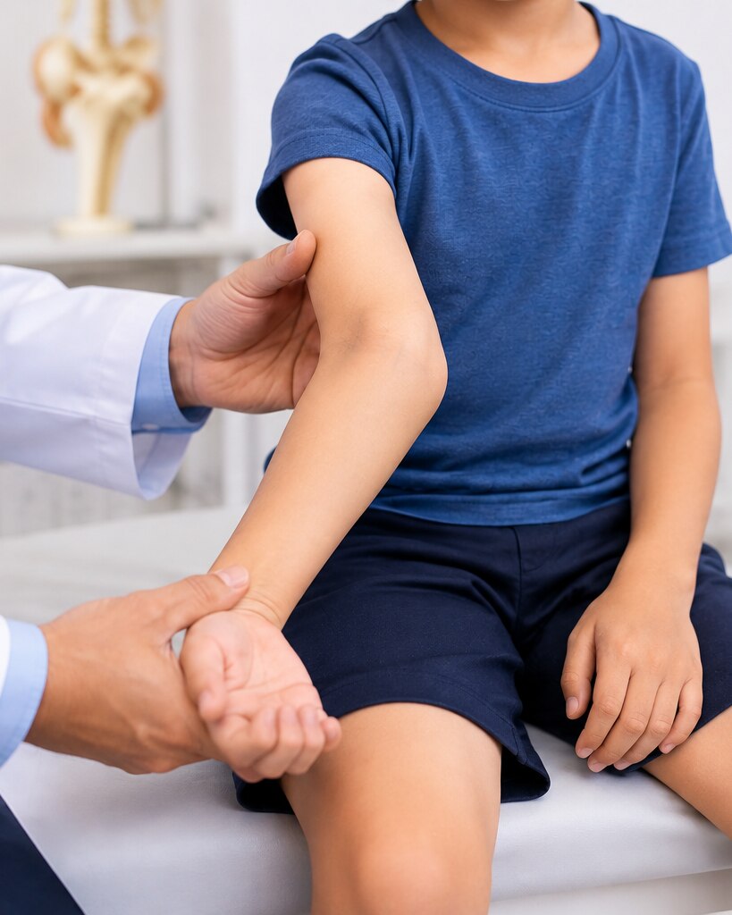
Clinicians measure it with both elbows extended and forearms facing upward, then compare the two sides; appearance alone cannot confirm the deformity.
| Condition | What the angle means | Typical appearance |
|---|---|---|
| Cubitus valgus | Excessive outward angulation of the forearm | The forearm angles away from the body |
| Normal carrying angle | The usual small outward angle | The hand clears the hip during walking |
| Cubitus varus comparison | The forearm angles inward more than expected | The elbow may look like a “gunstock” deformity |
Cubitus valgus can develop because the outer part of the elbow grows unevenly or fails to heal normally. Recognised causes include:
- A congenital elbow deformity or a bone-development condition present from early childhood
- A lateral condyle fracture that heals in poor alignment, or a non-union that disrupts elbow growth
- A growth-plate injury after an elbow fracture; supracondylar fracture healing can also alter alignment, although it more often produces cubitus varus
- Skeletal dysplasia or chromosomal conditions such as Turner syndrome
The injury’s type, location, and timing matter because correcting only the visible angle can miss an unstable or unhealed lateral condyle.
Which Symptoms Need an Examination, and How Is the Deformity Confirmed?
An examination is warranted when cubitus valgus symptoms include progressive outward angling, elbow instability, pain, reduced movement, or weakness. Ring and little finger numbness, tingling, hand weakness, or wasting of the small hand muscles point to pressure or traction on the ulnar nerve and possible tardy ulnar nerve palsy.
Cosmetic asymmetry alone does not prove a clinically important deformity.
| Finding | What it may indicate | How it is assessed |
|---|---|---|
| Different elbow angle | Angular deformity or growth-related progression | Compare both arms with elbows straight |
| Numbness or weakness | Ulnar nerve involvement | Test sensation and finger-spreading strength |
| Pain or looseness | Instability or an associated fracture problem | Check elbow stability and movement |
| Limited bending or straightening | Joint or bone abnormality | Measure the range of motion |
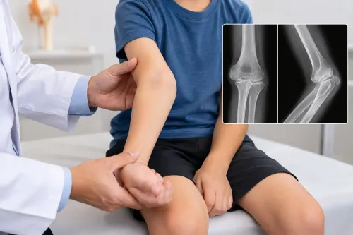
The clinician measures the carrying angle with both elbows extended and forearms turned palm-up, then compares the sides. The examination also records tenderness, skin changes, prior fracture effects, and ulnar-nerve motor and sensory function.
X-rays confirm the alignment rather than relying on appearance. An anteroposterior radiograph allows measurement of the humeroulnar angle and shows whether the deformity comes from the upper arm or forearm side of the joint.
Additional views can reveal a lateral condyle nonunion, malunion, growth-plate injury, instability, or another structural cause that requires more than simple angular correction.
When Is Observation Appropriate, and Can Exercises Correct the Angle?
Observation is appropriate for a mild, stable, painless deformity when elbow movement is preserved and there are no ulnar-nerve findings. The clinician establishes a baseline and checks for change over time rather than treating the appearance alone.
At each review, assessment focuses on:
- The carrying angle compared with the opposite elbow
- Elbow flexion, extension, and overall motion
- Instability or weakness during use
- Numbness, tingling, or weakness in the ring and little fingers
Follow-up X-rays are added when the deformity developed after an elbow injury, especially a lateral condyle fracture. They show whether alignment is changing, healing is incomplete, or growth-related progression is developing. A child with a previous lateral condyle non-union or malunion needs closer review because delayed ulnar-nerve symptoms can appear even after a painless period.
Exercises and physiotherapy do not usually straighten a fixed bony angle. Physiotherapy can preserve elbow motion, maintain strength, and address weakness caused by altered mechanics. Bracing supports the joint in selected situations but does not remodel an established deformity.
If the angle increases, instability develops, function declines, or nerve symptoms appear, observation is no longer enough. Treatment must address the structural cause; that may require reconstruction of a damaged lateral condyle or a corrective operation rather than exercises alone.
When Is Surgery Considered, and Which Corrective Operation Is Used?
Surgery is considered when the deformity is progressive, painful, unstable, functionally limiting, or associated with ulnar-nerve symptoms such as tingling, numbness, weakness, or muscle wasting.
A major cosmetic concern can justify discussion, but an operation is not automatically appropriate for an otherwise symptom-free child because correction carries risks of stiffness, infection, nonunion, hardware failure, nerve injury, and recurrence during growth.
The usual operation is a distal humeral corrective osteotomy. The choice depends on the bone shape, an old fracture’s malunion or nonunion, nerve findings, age, and remaining growth.
| Option | Main use | Growth and planning consideration |
|---|---|---|
| Lateral closing-wedge osteotomy | Removes a wedge from the outer distal humerus to reduce valgus | Reliable for a single-plane correction; growth-related recurrence must be considered |
| Medial opening-wedge osteotomy | Opens the inner side to change the distal-humerus angle | Requires stable fixation and careful assessment of bone quality and the ulnar nerve |
| Dome osteotomy | Corrects the angle through a curved cut, allowing adjustment in more than one plane | Useful for complex or multiplanar deformity |
| 3D-planned correction | Uses CT-based planning and guides for an irregular post-fracture shape | Helps match correction to the child’s anatomy rather than relying on one angle |
A younger child with substantial growth remaining needs a plan that accounts for later change; a near-mature adolescent may need a more definitive correction. If a lateral condyle fracture has not united, or healed in poor position, treating that structural problem may accompany the osteotomy.
Nerve compression strengthens the case for timely correction and possible ulnar-nerve release.
What Should Families Expect From Surgery, Recovery, and Specialist Review?
Surgery planning starts with confirming the deformity, elbow motion, stability, and ulnar-nerve function rather than correcting appearance alone. Bring old X-rays, fracture and operation reports, injury dates, growth or medical records, medication and allergy lists, and a note of numbness, weakness, or activity limits.
1. Before correction, the specialist compares both elbows, reviews current imaging, and explains the osteotomy level, fixation, and whether nerve decompression or anterior transposition is needed. A review at About The Bone & Joints - Advance Ortho Care should document small- and ring-finger sensation, hand strength, and muscle wasting before surgery.
2. After the bone is realigned, plates and screws usually hold the correction; a cast or brace protects it while healing begins. Elbow osteotomy recovery includes wound checks, repeat X-rays, gradual motion exercises, and later strengthening. Your surgeon sets the timing for school, lifting, and a safe return to sport.
3. Ask about loss of correction during growth, stiffness, infection, hardware irritation, nerve or blood-vessel injury, malunion, and delayed union and non-union. Persistent or worsening ulnar-nerve symptoms deserve prompt review because longstanding compression may not fully recover.
4. Seek urgent assessment for a cold, pale, blue, or rapidly swollen hand; new inability to move the fingers; severe escalating pain; fresh numbness or weakness; fever with wound drainage; or a new injury. Stable appearance concerns belong in routine specialist planning.
Related services
Paediatric orthopaedics treatment and management Paediatric orthopaedics treatment and management Dr. Darshan Shinagne, based on Kolshet Road, Thane, provides specialized paediatric orthopaedic care for children with bone, joint, and muscle... View service → |
Osteotomy and deformity correction Osteotomy and deformity correction Dr. Darshan Shinagne, located on Kolshet Road, Thane, provides advanced osteotomy surgery treatments for patients with joint deformities, arthritis,... View service → |
Frequently asked questions
What causes cubitus valgus in children?
Cubitus valgus can result from an elbow fracture that healed with altered alignment, a growth-plate injury, or less commonly a congenital or developmental difference.
Which symptoms need an examination for cubitus valgus?
Arrange an examination for a visibly increasing angle, elbow pain, weakness, reduced movement, hand muscle wasting, or numbness and tingling in the ring and little fingers.
Can exercises correct the cubitus valgus angle?
Exercises can maintain strength and movement but cannot straighten a fixed bony angle. Observation is appropriate when the deformity is mild, stable, painless, and causes no nerve or functional problems.
When is surgery used for cubitus valgus?
Surgery is considered when the deformity is substantial or progressive, causes ulnar-nerve symptoms or functional difficulty, or creates a significant appearance concern after specialist assessment.
What operation corrects cubitus valgus?
A corrective distal humerus osteotomy removes or reshapes a bone wedge and repositions the elbow, with fixation using plates, screws, or pins selected by the surgeon.