Cubitus varus changes the elbow’s carrying angle so the forearm points toward the body when the arm is straight, creating the appearance often called gunstock deformity. By the end, you will know how clinicians measure the alignment, when a painless deformity can be observed, and what corrective surgery involves when symptoms or mechanics justify it.
Key takeaways
- Cubitus varus commonly develops after a childhood supracondylar humerus fracture.
- Doctors compare both carrying angles and use X-rays to assess alignment and cause.
- Observation may suit a painless, stable deformity without functional or nerve problems.
- Corrective distal humeral osteotomy realigns the lower upper-arm bone when treatment is needed.
What cubitus varus changes—and which elbow problems can look similar
An inward-angulated elbow points the forearm toward the body when your arm is straight, rather than slightly away from it. This reduced or reversed carrying angle is called cubitus varus deformity, or gunstock deformity. It commonly follows a childhood supracondylar humerus fracture that healed with the lower humerus tilted inward.
The appearance may be cosmetic, but the deformity can also include rotation or front-to-back malalignment that a simple angle misses.
| Condition | What may look similar | Clue that separates it |
|---|---|---|
| Cubitus valgus | The elbow appears misaligned when the arm is extended | The forearm angles away from the body, increasing the carrying angle |
| Rotational or sagittal malunion | The elbow looks crooked despite a modest side-to-side angle | Turning the arm or bending it changes the appearance; motion may be restricted |
| Posterolateral rotatory instability | The elbow seems to shift or point abnormally | Giving-way, apprehension while pushing up, or recurrent slipping suggests instability |
Arrange an assessment if the difference between elbows is obvious, painful, worsening, or linked to reduced movement. Seek examination after an old fracture if you have:
- Elbow snapping, especially from the triceps
- Giving-way or a sense that the joint is unstable
- Numbness or tingling in the ring and little fingers
- Weak grip or hand clumsiness
- Difficulty straightening, bending, or rotating the forearm
A painless, stable asymmetry does not automatically require corrective surgery, but waiting for a healed fracture to remodel the carrying angle is unreliable.
How clinicians measure elbow alignment and find the cause
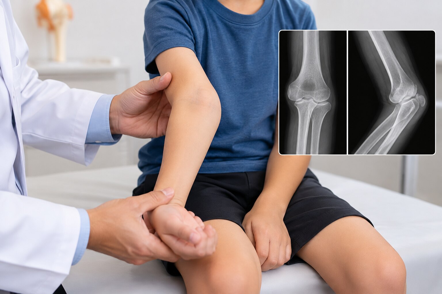
A clinician confirms cubitus varus by comparing both arms, not by judging one elbow against a population average. During elbow alignment assessment, the examiner checks the bilateral carrying angle with both arms extended, observes forearm rotation, measures motion, and looks for instability or ulnar-nerve irritation.
Standardized imaging provides the numerical comparison. Bilateral standing or standardized anteroposterior radiographs show whether the injured elbow differs from the opposite side. The carrying angle, also called the humeroulnar angle, describes the coronal-plane relationship between the upper arm and forearm. A Baumann angle radiograph helps assess distal-humerus tilt, especially after a childhood supracondylar fracture.
Position matters. Rotating the arm during an X-ray can substantially change the apparent Baumann angle and make the deformity look milder or worse than it is. The images must therefore be taken in a reproducible position and compared with the opposite elbow when possible.
The cause comes from the history and the shape of the healed bone. Old fracture records and earlier films can show whether the distal humerus healed in varus; current images can reveal growth-related or post-traumatic malalignment. A simple carrying-angle difference mainly reflects coronal angulation.
If the distal humerus also has rotational or flexion-extension malalignment, the elbow has a three-dimensional deformity that ordinary measurements may miss. CT or three-dimensional planning is considered when that detail affects reconstruction.
When observation is reasonable—and when the deformity becomes a treatment problem
A stable, painless cubitus varus usually needs no immediate operation when motion, strength, and elbow stability are normal and the appearance does not trouble the patient. After a childhood supracondylar humerus fracture, a distal humerus malunion will not reliably remodel to a normal carrying angle, so observation means monitoring—not waiting for dependable spontaneous correction.
Correction becomes worth discussing when the deformity affects daily use, sport, clothing, or self-confidence, or when examination shows mechanical or nerve problems. The decision depends on symptoms, skeletal maturity, deformity magnitude, three-dimensional shape on radiographs, and the patient’s goals; no single angle mandates surgery.
- The appearance remains unacceptable to the patient after the trade-off is explained.
- Reduced motion, weakness, recurrent instability, or progressive mechanical symptoms develops.
- Snapping triceps or suspected posterolateral rotatory instability changes elbow function.
- Ulnar nerve symptoms, such as numbness or tingling in the ring and little fingers, hand weakness, or inner-elbow pain, appear.
- Imaging shows substantial rotational or sagittal malalignment that the ordinary carrying-angle measurement misses.
Corrective osteotomy is not a cosmetic reset. Risks include nerve injury, infection, delayed union or nonunion, stiffness, hardware irritation, undercorrection, recurrence, and overcorrection into valgus. Discussion is strongest when the expected functional or personal benefit clearly outweighs those risks.
What corrective distal humeral osteotomy changes and how techniques differ
Corrective osteotomy restores elbow alignment by cutting the distal humerus, repositioning its lower segment, and fixing it so the forearm points at a more appropriate carrying angle. The correction can address varus angulation alone or combine angular, rotational, and translational changes when three-dimensional malalignment is present.
| Technique | What it changes | Why choose it or avoid it |
|---|---|---|
| Lateral closing-wedge osteotomy | Removes a bone wedge and closes the outer side | Straightforward and commonly used, but it can leave a lateral bony prominence and offers less control of rotation or translation |
| Dome osteotomy | Rotates the distal fragment around a curved cut | Useful when the surgeon needs gradual angular correction with some rotational adjustment; the curved cut requires precise planning and fixation |
| Step-cut osteotomy | Uses interlocking cuts to reposition the bone | Can provide angular and rotational correction with useful bony contact, but the operation is technically more demanding |
| Opening-wedge or three-dimensional osteotomy | Opens one side or corrects several planes together | Suits selected deformities needing translation, rotation, or length adjustment; it may require more fixation, grafting, and healing time |
The surgeon chooses the method from the size and direction of the deformity, the amount of rotation or translation, bone quality, growth status, and the need to avoid nerves and joint stiffness.
A simple lateral closing-wedge osteotomy may fit a mild, mainly angular deformity; a complex three-dimensional correction can better match multiplanar malalignment but carries greater technical and healing demands. Even a well-planned operation can leave undercorrection, overcorrection, or recurrent deformity.
Planning, recovery, alternatives, and questions to take to consultation
A clear operation has a measurable goal: restore the planned alignment, improve stability or mechanics, and address a defined nerve problem—not simply make the elbow look straighter. Preoperative planning compares both arms, reviews radiographs, and uses CT for elbow deformity when rotational or three-dimensional malalignment is suspected.
Before surgery, ask how the correction will be measured and whether the plan uses plate and screw fixation, another fixation method, or staged treatment. Afterward, expect protection in a splint or brace, wound checks, repeat radiographs, and gradual motion exercises.
Bone healing determines when lifting and sport can resume; stiffness is a common trade-off, so rehabilitation matters.
Discuss these decisions directly:
- What symptom or mechanical problem is the operation expected to improve, and how will success be measured?
- Is observation reasonable if the deformity is painless and stable? Compare that choice with surgery, especially when the concern is cosmetic.
- What are the risks of nerve injury, infection, delayed union, nonunion, hardware irritation, recurrence, undercorrection, overcorrection into valgus, and stiffness?
- If you have numbness in the ring or small finger, hand weakness, or a snapping nerve, is ulnar nerve decompression or transposition needed? Alignment correction will not reliably reverse established nerve damage.
- Could instability, snapping triceps, or another mechanical problem require a different procedure?
About The Bone & Joints - Advance Ortho Care can help structure an elbow consultation around imaging, nerve findings, fixation choice, and a specific outcome rather than an angle alone.
Related services
Osteotomy and deformity correction Osteotomy and deformity correction Dr. Darshan Shinagne, located on Kolshet Road, Thane, provides advanced osteotomy surgery treatments for patients with joint deformities, arthritis,... View service → |
Shoulder and Elbow Surgery Dr. Darshan Shinagne, practicing on Kolshet Road, Thane, provides expert shoulder and elbow surgery treatments for patients suffering from joint... View service → |
Frequently asked questions
What is cubitus varus deformity?
Cubitus varus is an inward-angulated elbow in which the forearm points toward the body when the arm is straight. It is also called gunstock deformity.
How do clinicians measure cubitus varus?
They compare the carrying angle of both elbows during examination and use X-rays to measure the deformity and assess how the humerus healed.
When does cubitus varus require treatment?
Treatment becomes a concern when the deformity causes functional limitations, pain, instability, nerve symptoms, progressive problems, or significant dissatisfaction with appearance.
What surgery corrects cubitus varus?
A corrective distal humeral osteotomy cuts and realigns the lower humerus, then holds the corrected position with fixation while the bone heals.