Developmental dysplasia of the hip can range from a mildly immature socket to a hip that is partly or completely dislocated, and the difference is not always visible at birth. Knowing which signs, risk factors, examinations, and age-appropriate scans matter helps you seek assessment before walking problems appear and understand why early treatment protects hip growth.
Key takeaways
- Ask about DDH screening before walking problems appear.
- Breech birth and family history increase the need for careful follow-up.
- Ultrasound helps assess infant hips before bones become clearly visible on X-rays.
- Early treatment can guide the hip socket and femoral head as they develop.
What DDH changes inside a child’s developing hip
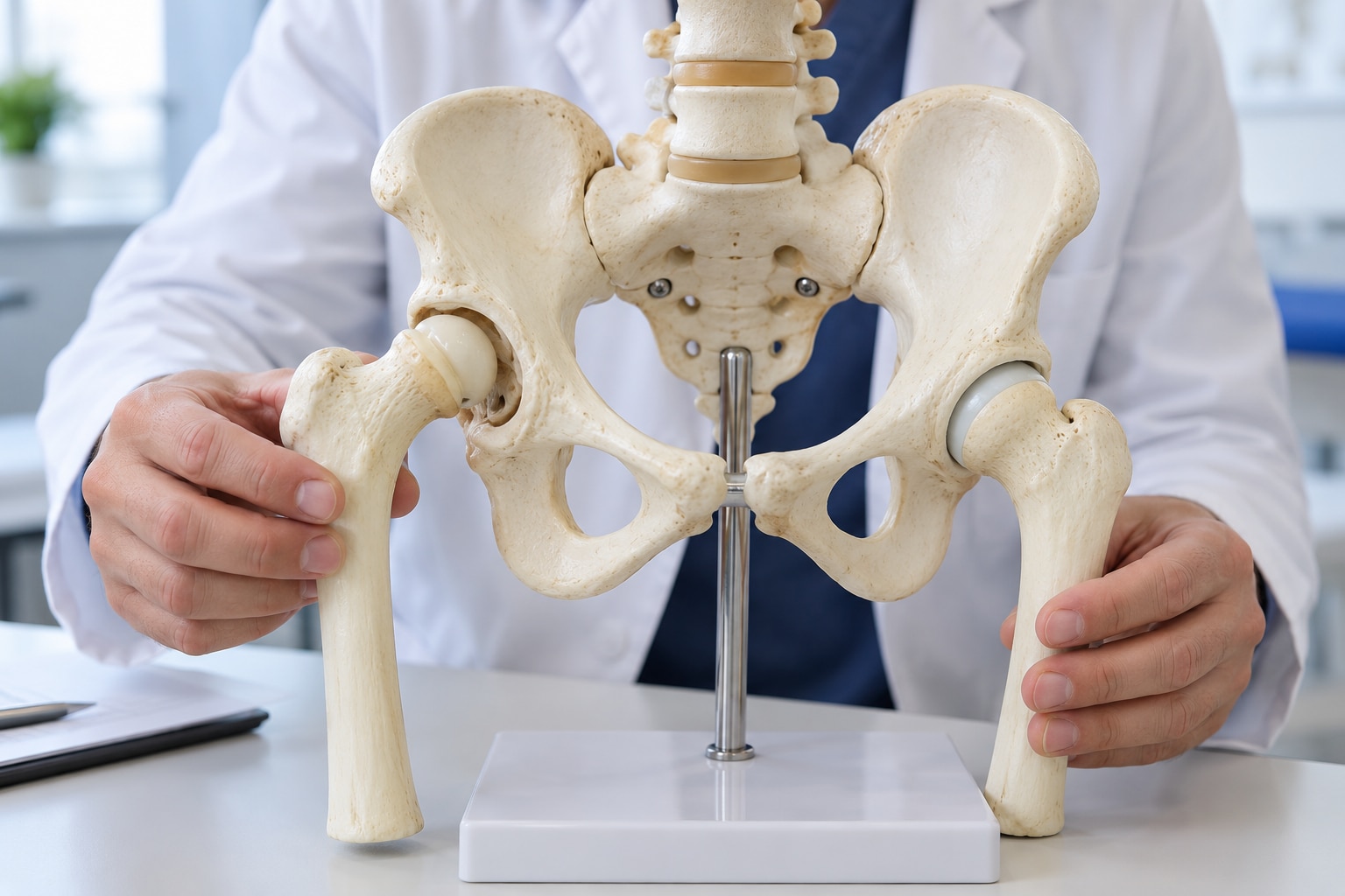
Developmental dysplasia of the hip (DDH) describes a hip joint that has not developed normally. The acetabulum, the socket in the pelvis, may be too shallow to hold the femoral head securely.
The hip may look normal but show femoral head instability, or the head may partly slip out of the socket (subluxation) or become completely dislocated.
A healthy hip keeps the femoral head centered while a child grows. Its contact and movement help deepen the acetabulum and shape the head into a smooth, rounded joint. If the head stays partly or completely displaced, that normal pressure is lost.
The socket can remain shallow, and the joint may become harder to position as the child gets older.
An untreated hip can cause:
- A limp, waddling gait, or difficulty walking because the leg cannot move through a stable joint.
- Unequal leg length when the femoral head sits higher or outside the socket.
- Hip, groin, or later joint pain as abnormal forces wear the joint.
Early DDH may cause no obvious discomfort, so waiting for walking problems delays care. A persistently displaced hip can require more invasive treatment later and raise the risk of early arthritis. Diagnosis and appropriate positioning during infancy give the femoral head the best opportunity to remain centered while the socket develops.
Which babies need careful hip screening and follow-up
A normal newborn hip examination does not rule out developmental dysplasia of the hip (DDH). Some instability appears later, while some abnormalities are difficult to detect by touch, so every baby needs repeated hip checks at routine visits.
Pay particular attention when a baby has:
- Breech presentation, especially if the baby was breech late in pregnancy or at delivery.
- A close family history of DDH.
- Female sex.
- First pregnancy, particularly when combined with breech position or low amniotic fluid (oligohydramnios).
- Tight swaddling that holds the legs straight and together; leave room for the hips to flex and the knees to move apart.
| Finding | What it can indicate | What to do |
|---|---|---|
| Limited movement when the thighs are gently moved apart | A restricted or poorly developing hip | Arrange prompt clinical assessment |
| Unequal thigh or buttock creases | Possible difference in hip position, though creases alone do not diagnose DDH | Mention it at the next visit or request earlier review if other signs exist |
| One leg appearing shorter | A displaced or poorly centered hip | Seek assessment promptly |
| A click or hip that feels unstable during examination | Hip looseness, subluxation, or dislocation | Let a clinician examine it; do not test repeatedly at home |
Risk factors raise suspicion but are not required. A baby without any known risk factor still needs surveillance, and an abnormal examination deserves assessment even when the baby feeds, sleeps, and moves normally.
How examination and imaging confirm DDH at different ages
A screening examination asks whether a baby needs closer attention; a diagnosis combines examination findings with age-appropriate imaging to define instability, subluxation, or dislocation. A normal newborn check lowers concern but does not rule out DDH, so clinicians reassess the hips at later well-child visits.
In young infants, clinicians examine both hips for:
- A palpable clunk during the Barlow maneuver, which suggests the femoral head can be displaced
- A clunk during the Ortolani maneuver, which suggests a displaced head can be reduced
- Limited hip abduction, unequal leg length, or asymmetric thigh and buttock creases
A soft click alone does not confirm disease. A persistently unstable hip, restricted movement, or an equivocal examination calls for imaging rather than a diagnosis based on screening alone.
- From birth to about 4 to 6 months, a hip ultrasound shows the cartilage-based femoral head and socket. It is appropriate for an abnormal or uncertain examination and for infants with important risk factors, but a mildly immature ultrasound does not automatically mean treatment; repeat examination or imaging may show normal development.
- After about 4 to 6 months, an AP pelvis X-ray becomes more useful as bone formation makes the femoral head and socket visible. It helps assess alignment and socket development when examination remains abnormal.
After walking begins, a limp, leg-length difference, or waddling gait can reveal a missed problem even when infant maneuvers are no longer informative.
How treatment supports hip development from infancy through childhood
Treatment is least invasive when the hip is found early. In the first months, a Pavlik harness holds the hips bent and gently apart so the femoral head stays centered in the socket while both structures develop.
It must not force the legs straight, and caregivers should not alter its straps without the treating team’s instructions.
Follow-up ultrasound checks whether the femoral head remains reduced and identifies skin, nerve, or positioning problems. If the harness fails or the diagnosis comes later, a clinician may use closed reduction under anaesthesia, then a spica cast to hold the hip in position while the socket develops.
After roughly 6 to 18 months, or when closed reduction cannot maintain alignment, open reduction may be needed to place the femoral head into the socket directly. Older children may also need a femoral or pelvic osteotomy to improve the socket or bone alignment.
These operations bring longer immobilisation and greater treatment burden than successful early harness treatment.
Keep every examination and imaging appointment; the hip can slip out again without obvious pain. Seek prompt clinical advice if you notice:
- A swollen, blistered, or persistently reddened area under the harness or cast
- A cold, pale, blue, or unusually swollen foot
- Reduced leg movement, weak kicking, or a new limp
- Apparent leg-length difference or worsening difficulty moving one hip
- Fever, foul odour, or a cast that becomes wet, cracked, or loose
What parents should do instead of waiting for walking problems
Request a review before walking if an infant has limited hip abduction, leg-length or crease asymmetry, a palpable clunk, breech presentation, close family history, or another clinician has raised concern. Once walking starts, do not wait for an abnormal gait, limp, waddling, or apparent leg-length difference; arrange assessment promptly.
Safe swaddling and correctly used baby carriers support healthy positioning, but they do not diagnose or correct an unstable hip. Keep the knees bent and apart, avoid wrapping the legs straight and tightly together, and never adjust a harness without the treating team’s instructions. These measures cannot replace examination, age-appropriate imaging, or follow-up imaging.
| Action | What it can answer | What it cannot replace |
|---|---|---|
| Home positioning | Reduces harmful tight positioning | Clinical assessment |
| Infant ultrasound | Assesses cartilage-rich hips under 4–6 months | Examination or review of symptoms |
| AP pelvis radiograph | Gives more useful bone detail after about 4–6 months | Specialist interpretation and follow-up |
- Is the hip stable, mildly immature, subluxated, or dislocated?
- Which imaging test fits my child’s age, and when is follow-up imaging needed?
- If observation or a harness is chosen, what positioning, skin checks, duration, and warning signs apply?
- What finding means we should return sooner?
About The Bone & Joints - Advance Ortho Care can help families organise these age-specific questions during a paediatric orthopaedic review in Thane.
Related service
Paediatric orthopaedics treatment and management Paediatric orthopaedics treatment and management Dr. Darshan Shinagne, based on Kolshet Road, Thane, provides specialized paediatric orthopaedic care for children with bone, joint, and muscle... View service → |
Frequently asked questions
What changes inside a child’s hip with developmental dysplasia?
The acetabulum may be too shallow to hold the femoral head securely, allowing the hip to become unstable or dislocated as it develops.
Which babies need careful DDH screening and follow-up?
Babies born breech and children with a family history of DDH need careful screening and follow-up. A limited hip examination or concerning findings also require further assessment.
How do doctors confirm DDH at different ages?
Doctors use a physical hip examination first. Ultrasound is useful in young infants, while an X-ray becomes more informative as the bones develop.
How is DDH treated from infancy through childhood?
Treatment depends on the child’s age and hip stability. It can include positioning in a harness, reduction and casting, or surgery when earlier treatment does not restore hip development.
What should parents do instead of waiting for walking problems?
Arrange a hip examination when risk factors or concerns are present, attend recommended imaging appointments, and seek review for limited hip movement or unequal leg length.