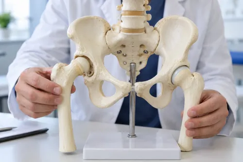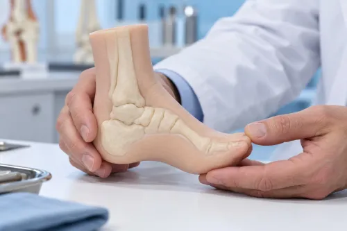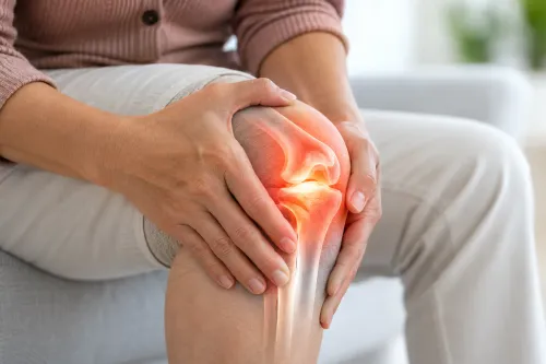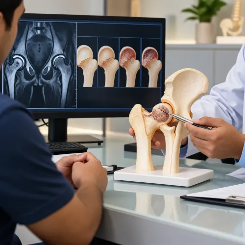
A child with Perthes disease may first show only a painless limp or complain of knee pain, while the problem is developing in the hip. You will learn which signs need assessment, why the condition occurs, how doctors confirm it, and how treatment protects hip shape during recovery.
Key takeaways
- Persistent limping, hip pain, or referred knee pain needs medical assessment.
- Perthes disease can flatten the femoral head after its blood supply is interrupted.
- Treatment protects hip shape through activity changes, physiotherapy, and specialist monitoring.
- Earlier diagnosis helps guide treatment before the femoral head loses more shape.
What Perthes Disease Is and Which Children Are Most Affected
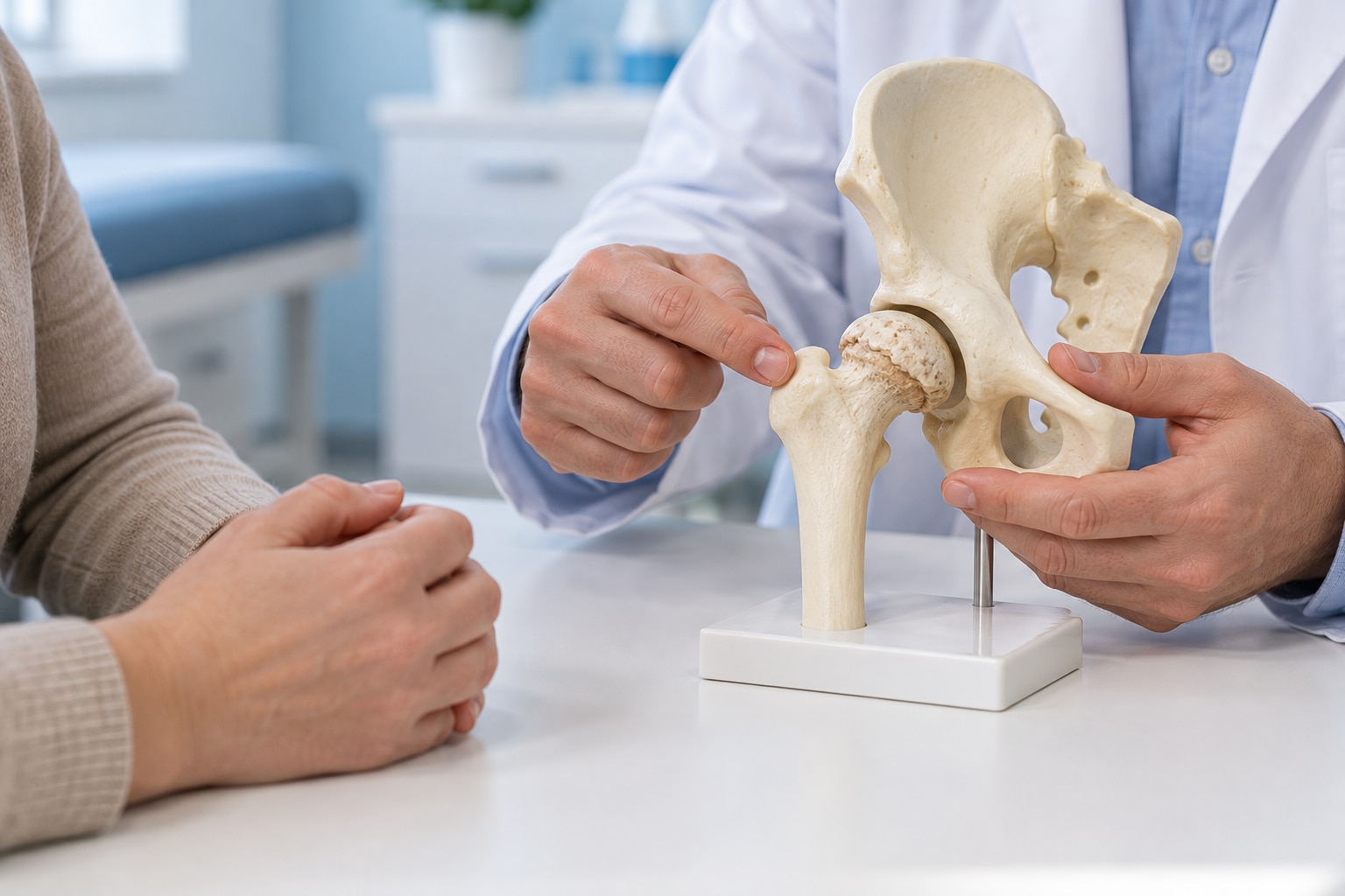
The hip’s ball-shaped growth area can lose its blood supply in Perthes disease. This avascular necrosis of the femoral head begins with a temporary interruption of blood flow, which weakens the bone, allows the femoral head to flatten or collapse, and then triggers gradual repair and reshaping.
The cause is usually unknown; do not routinely blame one injury, sports activity, or normal play.
| Pattern | What parents should know | What it means |
|---|---|---|
| Age | Most cases affect children aged 4 to 10 years | Age helps doctors estimate the hip’s ability to remodel |
| Sex | Boys are affected about four times as often as girls | A girl with a persistent limp still needs assessment |
| One or both hips | One hip is usually involved; both hips can develop disease at different times | A normal-looking opposite hip does not rule out Perthes disease |
| Possible associations | Low birth weight, delayed skeletal maturity, and household smoke exposure have been linked in research | These are associations, not proven single causes |
Perthes disease is not contagious and is not a result of poor parenting. A family history can occur, but most children have no clear inherited explanation.
Symptoms also do not predict the final hip shape: treatment decisions depend on the child’s age, disease stage, femoral-head involvement, hip movement, and whether the ball remains seated inside the socket.
Early Symptoms That Can Point to a Hip Problem
A painless limp is often the first clue, especially when a child is not complaining about pain. The limp can fluctuate over weeks or months and become more noticeable after running, climbing stairs, or sports. A child may shorten the stride, avoid putting weight on one side, or sit out of activities without explaining why.
Pain from the hip does not always feel like hip pain in children. Ask where the child points, and watch for:
- groin pain or aching at the front of the thigh
- referred knee pain, particularly when the knee looks normal
- stiffness after rest or difficulty spreading the legs apart
- pain or limping that worsens with activity
- reluctance to run, climb, or bear weight on one side
Knee pain deserves a hip assessment when examination of the knee finds no clear cause. Reduced hip movement, especially difficulty rotating the leg inward or moving it out to the side, can appear before an X-ray shows an obvious change. A normal early X-ray therefore does not settle the question if the limp persists.
Seek urgent medical care for sudden severe pain, fever, marked illness, or refusal to bear weight. Those features can indicate infection or another emergency rather than the slower pattern parents often notice first.
When a Limp Needs Prompt Assessment and How Perthes Is Diagnosed
Seek same-day urgent care if your child has sudden severe hip or leg pain, fever, looks seriously unwell, or refuses to bear weight. Septic arthritis and osteomyelitis can damage bone or the joint quickly; clinicians may order blood tests such as C-reactive protein and erythrocyte sedimentation rate, an ultrasound, or joint aspiration.
Arrange prompt assessment for a limp that persists, returns over several weeks, or comes with groin, thigh, or knee pain. A normal knee examination does not exclude a hip problem. The clinician compares both hips and checks:
- Reduced hip abduction, meaning difficulty moving the leg out to the side
- Limited internal rotation, often an early and important finding
- Pain, stiffness, leg-length difference, and the child’s walking pattern
Plain X-rays of both hips are usually the starting point, but a normal early X-ray does not rule out Perthes disease. Repeat radiographs may reveal changes later. MRI for Perthes disease detects early bone and blood-supply changes and helps when symptoms and examination findings remain suspicious.
| Test or finding | What it helps distinguish | Important limitation |
|---|---|---|
| Hip examination | Restricted motion suggests a hip source rather than an isolated knee problem | A mild early case can have subtle findings |
| Pelvis and hip X-rays | Bone shape, collapse, or another structural cause | Can be normal early |
| MRI | Early Perthes changes, marrow injury, or other hip disease | Use when specialist assessment justifies it |
| Blood tests, ultrasound, or aspiration | Infection or significant inflammation | Normal results do not replace hip imaging |
About The Bone & Joints - Advance Ortho Care can provide the specialist examination needed when a child’s limp and imaging do not match.
Related service
Paediatric orthopaedics treatment and management Paediatric orthopaedics treatment and management Dr. Darshan Shinagne, based on Kolshet Road, Thane, provides specialized paediatric orthopaedic care for children with bone, joint, and muscle... View service → |
How the Disease Progresses and How Treatment Protects the Hip
Over several years, the femoral head passes through collapse and fragmentation, followed by reossification and remodelling. The softened ball can flatten or move partly outside the acetabulum during this vulnerable period, then gradually rebuild. Pain and limping can improve before the bone has finished reshaping, so symptom relief does not confirm structural recovery.
Treatment depends on age, disease stage, the amount of femoral-head involvement, hip movement, and containment—not on the diagnosis alone.
| Option | Main approach | When it applies |
|---|---|---|
| Observation | Scheduled examinations and radiographs track containment, shape, motion, and leg-length differences | Younger children or limited disease |
| Brace-based containment treatment | An abduction brace keeps the head deeply seated while it heals | A child with usable motion and a head that can remain contained |
| Surgery | A femoral or pelvic osteotomy redirects the hip to maintain containment | Older children, substantial involvement, extrusion, or lost containment |
Hip movement is a treatment signal. Reduced abduction, or a head that cannot stay centred, increases the risk of a poor shape after healing; restoring and preserving motion therefore matters during every stage. Stretching, activity changes, physiotherapy, or a brace may protect movement, based on the treating team’s assessment.
Surgery is not automatically better than observation. Doctors weigh expected remodelling against operative risks and prolonged rehabilitation, with containment most valuable while the head remains soft. Even children managed without surgery need active surveillance until growth and remodelling show how the hip has settled.
Follow-Up, Recovery, and Long-Term Hip Outlook
Recovery can take several years because the femoral head passes through collapse, fragmentation, and reossification before its final shape is clear. Pain and limping can improve while reshaping continues, so symptom relief does not prove that the hip has recovered structurally.
Follow-up protects against missing loss of containment or worsening stiffness. Depending on age, disease stage, and hip movement, clinicians may use observation, physiotherapy, activity adjustments, bracing, or surgery. Younger children with limited involvement may need active surveillance rather than an operation, but surveillance includes scheduled radiographs and repeated examinations.
At each review, ask whether the following are being tracked:
- Hip range of motion, especially abduction and rotation
- Whether the femoral head remains contained inside the acetabulum
- Femoral-head shape as collapse and regrowth progress
- Hip congruence, meaning how smoothly the ball fits the socket
- Leg-length difference and growth-related changes
- Pain, limp, walking endurance, and daily function
The long-term outlook depends chiefly on the child’s age at disease onset, the amount of femoral-head involvement, the duration of containment, and the final relationship between the femoral head and acetabulum. A round, well-contained head that matches the socket has the best chance of remaining functional.
A flattened or poorly matched hip can leave restricted movement, a persistent limp, or leg-length discrepancy. It also increases the risk of early degenerative arthritis, which can cause pain and activity limits in adulthood. Keep appointments even after walking looks normal; the final X-ray shape matters more than early improvement.
Frequently asked questions
What are the early symptoms of Perthes disease?
Early symptoms include a painless limp, hip stiffness, groin or thigh pain, and pain referred to the knee.
When does a child’s limp need prompt assessment?
Arrange prompt assessment for a limp that persists, worsens, follows no clear injury, or occurs with hip or knee pain.
How is Perthes disease diagnosed?
A clinician assesses walking pattern and hip movement, then uses hip X-rays and further imaging when needed to evaluate the femoral head.
How is Perthes disease treated?
Treatment protects the hip by preserving movement, limiting impact activities, managing pain, using physiotherapy, and considering surgery when hip containment is at risk.
What is the long-term outlook for children with Perthes disease?
Recovery takes several years as the femoral head repairs and reshapes; the final hip outlook depends on age, disease severity, and retained hip shape.