A clubfoot is not simply a newborn’s foot rotated inward: it combines four changes in the arch, forefoot, heel, and ankle that require a precise examination and correction sequence. By the end, you will understand how clinicians confirm CTEV, what Ponseti casting and bracing involve, how relapse is managed, and which questions to ask before treatment begins.
Key takeaways
- CTEV combines inward turning, heel cord tightness, high arch, and forefoot deviation.
- Ponseti treatment uses gentle manipulation, weekly casts, and Achilles tenotomy when needed.
- A foot-abduction brace prevents relapse after casting and requires consistent family use.
- Long-term reviews detect stiffness, recurrent deformity, and brace-related problems early.
What CTEV looks like and how clinicians confirm the diagnosis
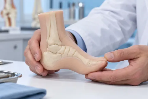
True congenital talipes equinovarus is a three-dimensional deformity, not simply a foot rotated inward. The four features of CTEV are:
- Cavus: an unusually high arch along the inner side of the foot.
- Forefoot adduction: the front of the foot angles toward the midline.
- Hindfoot varus: the heel turns inward.
- Equinus: the ankle points downward and resists upward movement.
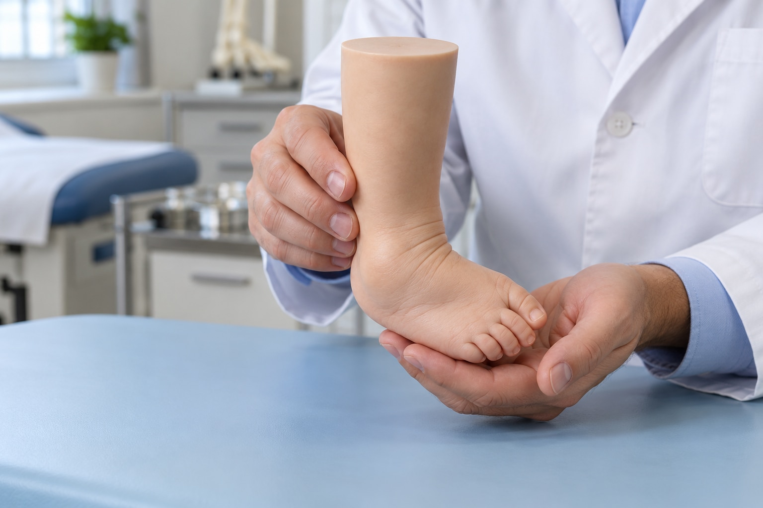
At birth, clinicians diagnose idiopathic clubfoot mainly by examination. They assess how rigid the foot is, the depth of its skin creases, calf size, and whether the ankle can be brought upward into dorsiflexion. A true CTEV foot remains curved and stiff when gently repositioned.
Other inward-turned feet have different examination findings:
- Positional talipes is usually flexible and corrects with gentle pressure.
- Metatarsus adductus affects mainly the forefoot; the heel and ankle are not fixed in the full clubfoot pattern.
- Vertical talus produces a rigid rocker-bottom shape rather than the high arch and inward heel of CTEV.
- Skew foot has a curved, complex alignment that does not match the classic four-part deformity.
- Neuromuscular or syndromic clubfoot may accompany weakness, stiffness in other joints, spina bifida, or arthrogryposis.
Prenatal ultrasound can suggest clubfoot, but the examination after birth confirms flexibility and whether it is isolated. X-rays are unnecessary in a typical newborn case; clinicians use imaging when the foot is atypical, rigid, recurrent, or linked to another skeletal condition.
What the examination reveals before treatment starts
At birth, the examination must establish whether this is a true clubfoot diagnosis, how flexible the foot is, and whether the infant has an isolated deformity. CTEV combines cavus, forefoot adduction, hindfoot varus, and equinus; a flexible positional talipes or metatarsus adductus follows a different treatment path.
1. Inspect the foot from above, behind, and underneath. Note the medial and posterior skin creases, arch height, heel position, forefoot alignment, calf size, and any difference between the two limbs.
2. Gently assess correction. The examiner checks whether the forefoot, heel, and ankle can be moved toward a plantigrade position, records ankle dorsiflexion, and identifies a rigid or severe deformity that could indicate atypical clubfoot.
3. Examine the whole infant, not only the feet. Check hip stability, knee movement, leg length, spine and skin over the lower back, muscle tone, limb formation, and joint flexibility. Findings such as hip instability, a spinal abnormality, weakness, or multiple stiff joints raise concern for spina bifida, neuromuscular disease, or arthrogryposis.
4. Decide whether tests are needed. Routine X-rays rarely clarify an uncomplicated newborn foot because much of it remains cartilage; imaging becomes useful for an atypical, rigid, recurrent, or suspected skeletal deformity. The recorded baseline helps judge correction by function—a painless, flexible, plantigrade foot that fits ordinary shoes—not by perfect visual symmetry.
How the Ponseti method corrects the foot step by step
Correction follows a sequence; forcing the entire foot outward at once can create a midfoot break and leave the heel turned in. The Ponseti method gradually reshapes the soft tissues while preserving movement in the joints.
1. The clinician gently manipulates the foot into the position that corrects cavus first. Elevating the first metatarsal aligns the forefoot with the hindfoot instead of bending the midfoot.
2. The foot is then gradually abducted around the head of the talus to correct forefoot adduction and hindfoot varus. Each position is held in an above-knee plaster cast, with the knee bent to keep the cast secure. Ponseti casting is usually repeated about once a week, and many infants need 4 to 8 casts.
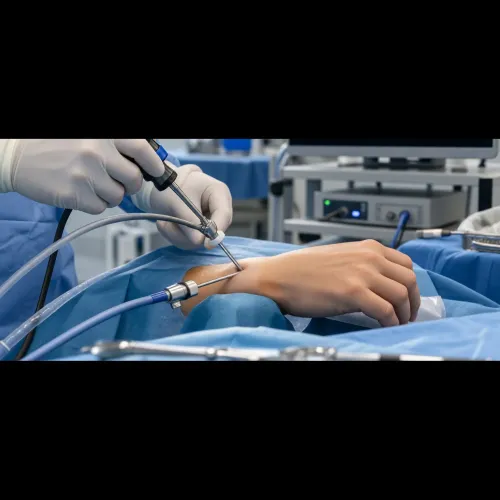
3. Equinus is corrected last because the tight Achilles tendon limits upward ankle movement. After cavus, adduction, and varus have improved, persistent equinus usually requires a percutaneous Achilles tenotomy rather than forcing the ankle upward inside a cast. This brief procedure is followed by another cast.
4. The final cast holds the foot in its corrected position while the tenotomy heals and the ankle gains dorsiflexion. It usually remains for about 3 weeks, allowing the tendon to heal at the new length and completing the initial correction.
The number of casts varies with severity and technique. A difficult correction or unexpectedly high cast count calls for reassessment for atypical or resistant clubfoot.
Why bracing and long-term follow-up determine the result
After the final cast comes off, the foot-abduction brace protects the correction while the child grows. Families usually use it for about 23 hours a day for roughly three months, then during sleep and naps until around age 4 or 5. The orthopaedic team sets the exact schedule.
Brace adherence matters: missed nights allow the tightened tissues to pull the foot inward again.
A clubfoot relapse can appear even after an initially excellent correction, so follow-up must continue for years. Watch for the foot turning inward, the heel lifting, reduced ankle dorsiflexion, or the child walking on the outer edge.
A child who starts walking with the front of the foot angled inward may have dynamic supination, in which the tibialis anterior overpulls the foot during gait. Report these changes promptly rather than waiting for the next routine visit.
Early relapse is not automatically a reason for extensive open surgery. The clinician can often restore flexibility with repeat manipulation and casting, followed by a repeat Achilles tenotomy when equinus has returned.
In a selected older child, tibialis anterior tendon transfer may be used after the foot has been passively corrected to address persistent dynamic supination. Continued examinations determine whether the brace needs adjustment, whether casting is enough, or whether surgery is appropriate.
Choosing a treatment plan and preparing for the family commitment
Ponseti treatment is usually the first choice for idiopathic clubfoot because it preserves movement and avoids the stiffness that can follow extensive surgery. It is a family treatment, not a single procedure: expect weekly cast visits, possible Achilles tenotomy, a final cast, and reliable brace use.
| Option | Main advantage | Main limitation |
|---|---|---|
| Ponseti method | Corrects most newborn feet with limited surgery | Relapse follows missed casting or bracing |
| Extensive corrective surgery | Addresses resistant deformity in one operative plan | Can leave the foot stiff, weak, or painful |
| Modified Ponseti treatment | Extends correction to older or complex feet | Needs more casts, closer review, and often has greater relapse risk |
Ponseti is incomplete when the foot remains pointed downward or turned inward after casting, the heel stays small and tight, or the required tenotomy and bracing plan has not been completed.
An atypical clubfoot, syndromic clubfoot, or neglected clubfoot may need modified manipulation, extra casts, imaging, treatment of an associated neurologic or muscular condition, or later deformity correction. Surgery is usually reserved for persistent or repeatedly relapsing deformity.
Before starting, ask your orthopaedic team:
- Is this idiopathic clubfoot, or are hip, spine, nerve, muscle, or limb findings present?
- How many casts do you expect, and what would an unusually high number suggest?
- Will a tenotomy be needed, and where will it occur?
- What brace schedule, fitting checks, and relapse signs should we follow?
- Who will manage the foot if correction remains incomplete?
About The Bone & Joints - Advance Ortho Care can help families obtain a second assessment when the diagnosis, correction, or need for surgery is uncertain.
Related service
Paediatric orthopaedics treatment and management Paediatric orthopaedics treatment and management Dr. Darshan Shinagne, based on Kolshet Road, Thane, provides specialized paediatric orthopaedic care for children with bone, joint, and muscle... View service → |
Frequently asked questions
What does congenital talipes equinovarus look like?
CTEV is a three-dimensional deformity with cavus, adductus, varus, and equinus: a high arch, inward-pointing forefoot, turned-in heel, and downward-pointing ankle.
How do clinicians confirm a CTEV diagnosis?
Clinicians examine the foot’s flexibility, skin creases, calf size, pulses, movement, and the position of the heel and forefoot. Typical idiopathic CTEV is diagnosed clinically rather than through routine X-rays.
How does the Ponseti method correct clubfoot?
The clinician gently manipulates the foot and applies a series of casts, correcting cavus first, then adductus and varus, followed by equinus. A percutaneous Achilles tenotomy is used when ankle dorsiflexion remains limited.
Why is bracing important after Ponseti treatment?
The foot-abduction brace holds the corrected position while tissues mature. Consistent use during the prescribed schedule reduces the risk of recurrent clubfoot, and follow-up checks identify relapse early.